Fibrous Dysplasia: Clinical Presentation and Common Symptoms Fibrous Dysplasia is a rare bone disorder that weakens bones. It replaces healthy tissue with fibrous material. This can cause bones to break more easily. It’s important to know about this bone disorder. Finding it early helps manage…
Fibrous Dysplasia: Clinical Presentation and Common Symptoms
Fibrous Dysplasia is a rare bone disorder that weakens bones. It replaces healthy tissue with fibrous material. This can cause bones to break more easily. It’s important to know about this bone disorder. Finding it early helps manage symptoms and prevent serious problems. Early detection leads to better care from doctors. This article will look at the different parts of Fibrous Dysplasia. We’ll discuss the treatment options to help patients. Understanding the condition is key to choosing the right treatment options.
Understanding the Pathophysiology of Fibrous Dysplasia
Fibrous dysplasia disrupts how our bones grow and stay strong. It happens when the normal bone development process is messed up at a cellular level. Instead of making healthy bone, the body creates a soft, disorganized fibrous tissue.
Genetic Mutations and Bone Development
The main cause is a genetic change in the GNAS gene. This change happens after a baby is conceived, so it’s not passed down from parents. It messes with the signals that tell cells how to develop during bone development.
This mutation stops bone-building cells, called osteoblasts, from growing up. They stay young and keep growing, making a mess of tissue. This biological error stops the body from making strong bone needed for support.
The Role of Fibrous Tissue in Bone Weakening
The fibrous tissue grows in the bone marrow, taking over healthy bone. This makes the bone look like a “ground-glass” on scans. The new tissue is soft and not mineralized, making the bone weak.
This weakness makes bones highly susceptible to deformities and breaks. The bone can bow or change shape over time. Understanding these cellular pathways helps doctors predict how the condition will progress.
Epidemiology and Prevalence in the United States
Researchers are studying this rare skeletal condition in the U.S. It’s hard to get exact numbers because it’s so rare. But, they’re learning a lot from what they do find.
This helps them improve how they diagnose and treat patients. It’s all about making things better for those affected.
Demographic Trends and Age of Onset
This skeletal condition usually starts early in life. Most kids or teens get diagnosed during their growth years.
The symptoms often show up when bones are changing a lot. This is why catching it early is so important. It helps kids stay healthy for the long run.
Geographic and Genetic Considerations
Studies show this skeletal condition doesn’t favor certain places in the U.S. It’s found pretty evenly everywhere.
It’s mostly caused by random genetic changes early on. These changes aren’t passed down from parents. So, it pops up randomly, making it tough for doctors to keep an eye on.
Clinical Presentation and Common Symptoms
The symptoms of this disorder vary based on where the bone is affected. Some people may not show any signs for years. Others might notice small changes that get worse over time. Finding the problem early is key to keeping bones healthy.
Bone Pain and Physical Deformities
Many patients first feel pain in a specific bone. This pain comes from the bone growing and changing shape. As time goes on, this can cause bones to look different, like being bent or swollen.
The bone’s structure changes, and fibrous tissue starts to replace the healthy bone. This makes the bone lose its shape and strength. People might see a limb that looks bent or a swelling that feels hard.
Pathological Fractures and Mobility Issues
The bone’s weakened state makes it more likely to break. Even a small injury can cause a fracture. These fractures are painful and need quick medical help to heal right.
Having too much fibrous tissue in the bone can make it hard to move. This makes everyday tasks harder. Doctors often suggest physical therapy to keep muscles strong and support the bone.
Neurological Complications in Craniofacial Cases
When the skull or facial bones are affected, things get more complicated. The bone growing can press on nerves, causing problems with feeling or movement. Vision and hearing loss are serious risks if the bone grows too close to these areas.
Dealing with these cases needs a team of doctors to avoid lasting damage. It’s important to watch the fibrous tissue closely with scans. Early action is crucial to keep nerves working and quality of life good.
The Spectrum of Fibrous Dysplasia Types
Fibrous Dysplasia shows different symptoms based on how many bones are affected. Doctors divide it into types to understand and manage it better.
Monostotic Fibrous Dysplasia
Monostotic Fibrous Dysplasia affects one bone. It usually starts in teens or early twenties. Often, it doesn’t cause symptoms for a long time.
Lesions can be in bones like the ribs, femur, tibia, and face bones. Even though they’re in one place, they need regular checks to avoid problems.
Polyostotic Fibrous Dysplasia
Polyostotic Fibrous Dysplasia affects more than one bone. It’s usually more serious and starts earlier than the single-bone version.
People with this type might have big bone problems and a higher chance of broken bones. Treating it needs a team effort to keep bones strong and moving well.
Syndromic Associations Including McCune-Albright Syndrome
Sometimes, Fibrous Dysplasia comes with other health issues, like McCune-Albright syndrome.
This syndrome has three main parts:
- Polyostotic skeletal lesions that cause bone pain and deformity.
- Café-au-lait skin pigmentation, often appearing as irregular patches.
- Endocrine hyperfunction, such as precocious puberty or thyroid issues.
Spotting these connections is crucial for doctors. It helps catch hormonal problems early, which can greatly improve life for those with this condition.
Diagnostic Methods and Imaging Techniques
Accurate diagnosis methods are key to spotting the unique bone changes of this condition. Doctors use many methods to check each lesion well and watch it over time.
Radiographic Features and X-ray Analysis
X-rays are often the first step. They show a “ground-glass” look, where bone is replaced by fibrous tissue.
Experts look for certain signs in these images, like:
- Well-defined borders of the affected bone area.
- Expansion of the bone cortex.
- Thinning of the outer bone shell.
- Variations in bone density across the lesion.
Advanced Imaging: CT Scans and MRI
When X-rays aren’t enough, doctors use CT scans. CT scans are great at showing the exact shape of a lesion and how likely it is to break.
MRI is used to see soft tissue involvement. It helps doctors tell the fibrous matrix apart from healthy tissue, which is key for surgery planning.
The Role of Bone Scintigraphy
Bone scintigraphy, or a bone scan, is a strong tool for finding metabolic activity in bones. By using a radioactive tracer, doctors can spot areas of high bone activity that might not show up on regular films.
This method is very helpful for patients with many lesions. It lets specialists see the disease’s full spread in the body. This ensures no hidden problems are missed during treatment.
Differential Diagnosis and Bone Tumor Considerations
Getting the right diagnosis is key to managing any skeletal condition. Many bone disorders look similar on X-rays. So, doctors use detailed diagnosis methods to make sure they’re treating the right thing. This helps avoid unnecessary treatments and clears up any confusion for the patient.
Distinguishing from Other Skeletal Conditions
Doctors compare symptoms to rule out other bone problems. This is important because the wrong treatment can harm more than help. They look at conditions like:
- Non-ossifying fibromas
- Paget’s disease of bone
- Osteofibrous dysplasia
- Enchondromas
- Giant cell tumors
By studying where and how the lesion looks, doctors can guess what it might be. Advanced scans show details that help tell these bone tumors apart from harmless growths. This careful method is the best way to be sure.
Identifying Potencial Malignant Transformation
Even though cancer is rare, it’s something doctors watch for closely. People with big or many lesions might need to see their doctor more often. This is to catch any changes in pain or size early.
Doctors look for signs that might mean the tumor is getting worse. These signs include:
- Sudden, localized increase in bone pain
- Rapid expansion of a previously stable lesion
- New soft tissue masses appearing near the bone
- Unexpected fractures in areas that were previously asymptomatic
By keeping a close eye on things, doctors can catch any changes quickly. Regular check-ups help track how the bone is doing. This is the best way to protect the patient’s health in the long run.
Impact on Bone Health and Structural Integrity
Persistent lesions can change how bones grow and function. These changes often disrupt bone development. This makes the skeleton more prone to stress and structural failure. Keeping bone health in check is key for a good quality of life.
Long-term Effects on Skeletal Development
Fibrous tissue replacing healthy marrow weakens bones. This makes them struggle to support the body’s weight. Over time, these areas may become misshapen, causing stress on healthy tissues.
Patients often face chronic issues that need ongoing medical care. Early intervention is critical to track lesion progress during growth. Without it, the skeleton’s structure may be permanently damaged.
Managing Secondary Complications
Keeping bone health in check requires tackling secondary complications. Limb length discrepancies can cause gait issues and pain. Doctors use orthotics or surgery to balance the skeleton’s load.
Joint instability is another big challenge. It affects daily mobility. To tackle this, patients may benefit from:
- Physical therapy to strengthen muscles around the joints.
- Regular imaging to track bone alignment shifts.
- Customized bracing for necessary support during activity.
- Pain management to reduce inflammation and improve comfort.
By tackling these issues early, patients can often keep their mobility. Consistent monitoring is the heart of effective long-term care for this condition.
Current Medical Treatment Options
Effective treatment options aim to stabilize bone health and ease pain. This condition affects bone turnover, so doctors focus on slowing it down. They want to improve life quality and lower the risk of future problems.

Pharmacological Interventions and Bisphosphonates
Bisphosphonates are a key treatment. They stop osteoclasts from breaking down bones. This helps reduce bone turnover and boost bone density.
Doctors choose these drugs based on how bad the bone damage is and what the patient needs. They’re not a cure but help keep bones strong. It’s important to check how well they’re working and adjust as needed.
Pain Management Strategies
Dealing with chronic pain is a big part of treatment. Doctors use medicines and lifestyle changes to keep patients active and pain-free. These treatment options help with both sudden and ongoing pain.
Good pain management needs a team effort. Patients should work with their doctors to find the right plan. Common methods include:
- Non-steroidal anti-inflammatory drugs (NSAIDs) to reduce inflammation.
- Physical therapy to strengthen muscles around the weak bone.
- Supportive bracing to stabilize and lessen stress on weak spots.
- Changing activities to avoid movements that cause pain.
By using these different treatment options, patients can manage their condition better. Talking regularly with specialists helps make these plans work even better over time.
Surgical Approaches for Skeletal Correction
When other treatments don’t work, doctors use surgery to fix the skeleton. This is needed when fibrous tissue causes too much damage. Even though it’s not a bone tumor, it can harm bones a lot.
Curettage and Bone Grafting Procedures
Curettage aims to remove the bad tissue from the bone. Doctors scrape out the lesion to make room for new bone.
Then, they fill the space with a bone graft. This helps the bone grow back stronger. There are a few ways to do this:
- Autografts: Bone from the patient’s own body is used.
- Allografts: Bone from a donor is used.
- Synthetic substitutes: Special materials help the bone grow.
Internal Fixation and Stabilization Techniques
Internal fixation is key when bones are at risk of breaking. It gives the bone the support it needs to heal.
Doctors use special tools to hold the bone in place. This keeps the area stable while it heals. The benefits are:
- Stronger bones can bear more weight.
- It stops bones from breaking in high-stress areas.
- It helps the bones line up right.
Reconstructive Surgery for Craniofacial Deformities
Fixing the skull and face is very complex. Reconstructive surgery aims to fix looks and function.
It involves shaping the bone for better looks and function. Sometimes, it’s needed to relieve pressure on nerves or organs. Precision planning is key for good results.
Physical Therapy and Rehabilitation Strategies
Physical therapy is key for those with skeletal issues. A good rehab plan keeps bones strong and helps patients stay independent. With the help of therapists, people get plans that fit their needs.
Improving Range of Motion and Strength
The main aim of physical therapy is to boost mobility and protect bones. Therapists suggest low-impact exercises to strengthen muscles without harming bones. This muscle support is key for bone health.
Regular movement stops stiffness and keeps joints flexible. Patients should do controlled, repeated motions to build endurance. These exercises help stabilize the body and lower injury risks in daily life.
Adaptive Strategies for Daily Living
Living with a chronic bone condition means making smart changes to avoid injuries. Occupational therapists recommend using assistive devices to ease strain on weak limbs. These tools are essential for maintaining safety when doing everyday tasks at home or work.
Changing your home can also boost your life quality. Simple changes, like installing grab bars or using ergonomic furniture, help protect bone health by reducing fall risks. By making these adjustments, patients can stay confident and manage their condition well.
Living with a Chronic Bone Disorder
Dealing with a bone disorder can be tough, but having a strong support network helps a lot. Medical treatments help with the physical side, but the emotional journey is just as important. Acknowledging your feelings is the first step to managing it well over time.
Psychosocial Impact and Support Systems
Getting a diagnosis can bring up many emotions, like anxiety or frustration about physical limits. It’s key to know these feelings are normal when facing a chronic bone disorder. Talking to others who go through the same thing can offer comfort and useful tips.
Here are ways to boost your emotional health:
- Join patient advocacy groups to share experiences and coping strategies.
- See a counselor or therapist to deal with the emotional impact of the diagnosis.
- Talk openly with family and friends about what you need.
- Focus on activities that make you happy and reduce stress.
Navigating Healthcare and Specialist Care
Handling a complex bone disorder needs teamwork between your doctor and specialists. Being organized is key to keeping your treatment plan on track. You should be the main person in charge of your health info.
To make your medical care smoother, do these:
- Keep all your medical records and scans in one place, either in a binder or digitally.
- Write down questions for your doctors to make the most of your time with them.
- Make sure your whole care team knows about any new symptoms or changes in treatment.
- Get a second opinion if you’re unsure about your treatment plan.
Being proactive in your care can help you manage your condition better. Empowerment comes from knowledge and speaking up for your health needs. Remember, you’re the most important part of your healthcare team.
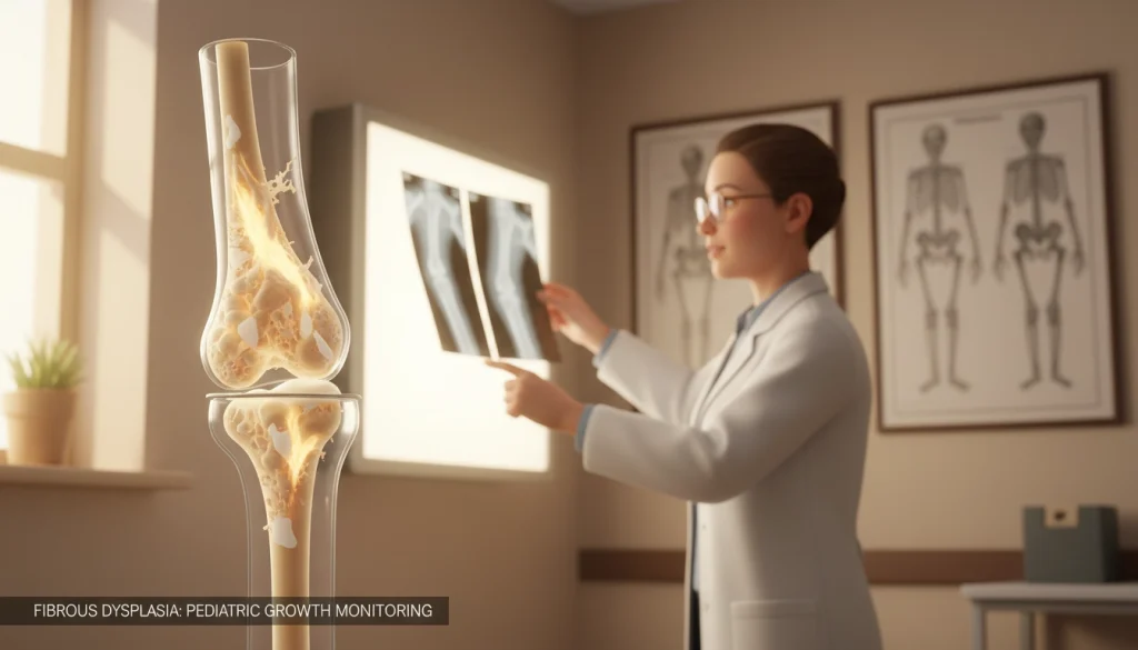
Pediatric Considerations and Growth Monitoring
It’s key to watch how this bone disorder affects kids as they grow. Kids change a lot physically, so they need special care. Doctors aim to help them grow while keeping their bones strong.
Managing the Condition During Childhood
Managing it well means checking the bones often. Doctors look at how the condition affects bone development. They use early steps to help kids move and feel better.
Parents and caregivers are very important. They notice if kids are more tired or in pain. Working with a team helps keep the treatment right for the child. This helps avoid problems with growing too fast.
Long-term Monitoring and Developmental Milestones
Watching how kids grow is a big part of their care. Doctors use these signs to see if the condition is slowing them down. They make changes to help kids reach their physical goals.
Doctors use scans and check-ups to see how bones are doing. This helps them plan for the future. Staying ahead helps kids live well into adulthood.
Emerging Research and Future Therapeutic Directions
Scientists are now focusing on the molecular roots of this disease to develop better care plans. Recent breakthroughs in genetic mapping have opened doors for more effective treatment options that go beyond traditional symptom management. This shift toward precision medicine offers hope for patients seeking long-term stability.
Targeted Therapies and Genetic Research
Current research centers on the GNAS gene mutation, which is the primary driver of the condition. By understanding how this mutation alters bone cell behavior, experts are developing targeted therapies designed to inhibit abnormal tissue growth at the source. These advancements aim to normalize bone development, not just react to damage.
Researchers are also exploring small-molecule inhibitors that could potentially halt the progression of fibrous lesions. These novel treatment options represent a significant leap forward in clinical science. By addressing the underlying genetic triggers, medical professionals hope to reduce the need for invasive interventions in the future.
Clinical Trials and Innovative Surgical Techniques
Ongoing clinical trials are currently evaluating the safety and efficacy of new pharmacological agents. These studies provide a vital pathway for patients to access cutting-edge care while contributing to global medical knowledge. Participation in these trials remains a cornerstone of progress for the entire patient community.
Beyond medication, surgeons are adopting innovative techniques to improve patient outcomes. The use of 3D-printed implants and computer-assisted navigation allows for more precise bone reconstruction. These modern treatment options minimize recovery time and enhance the structural integrity of affected areas, ensuring a better quality of life for those living with the condition.
Prognosis and Long-term Outlook for Patients
Managing Fibrous Dysplasia needs a proactive approach to keep bones healthy over time. Most people live well when they work closely with their doctors.
Regular visits to the doctor help track bone changes and catch problems early. This is the best way to avoid Fibrous Dysplasia complications.
Teams of doctors from different fields offer the best support. Orthopedics, endocrinology, and physical therapy specialists work together. They create plans tailored to each patient, helping manage symptoms and keep them mobile.
Living with Fibrous Dysplasia means adapting to physical needs. Many people do well by using modern medical tools and staying informed. Regular check-ups and talking openly with doctors help them stay active.
For more help and support, contact the Fibrous Dysplasia Foundation. Connecting with others who understand can offer great insight. Your journey is backed by ongoing medical research and dedicated care.
Clinical Expertise & Trust Center
Healthcare decisions often involve more than a single treatment option. The experts, technologies and centers presented here reflect areas of expertise that are commonly associated with this topic, helping patients better understand available care pathways across the Acibadem Healthcare Group network.